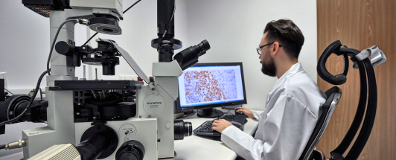
The aim of the project is to design and to implement of web-based platform for the computer analysis of microscopic images to support the pathological diagnosis. The platform will be free of charge and will offers to registered doctors, scientists and students:
- quantitative analysis of histological staining and tissues elected by algorithms branded by the team or the individual authors,
- archiving microscopic images,
- peer consultation, and join work independently from distance between scientific collaborating centers.
The use of proposed platform allows:
- to save pathologists time spend on quantitative analysis,
- to reduce consulting costs by replacing sending of the physical preparations by placing their images (mostly virtual slide) on the platform server,
- to increase reproducibility, comparability and objectivity of quantitative evaluations.
These effects carried out with a direct impact on improving the effectiveness and decreasing the costs of patients treatment.
It seems that offering pathomorphologists a tool aiding their work as a platform allowing access to digital microscopic image analysis system will be very helpful in executing time consuming quantitative analysis of the chosen immunohistemical stains as well as increase the credibility and comparability of results. Free access to the platform would lower the costs of a potential acquisition of individual systems of image analysis in separate diagnostic labs. What is more, the possibility of inserting an image of chosen fields of view or scanned whole slides in the database would allow consultation between the centers. A possibility of creating research groups working on the common database of disease cases is an important asset of the proposed platform. The history of analysis and consults will also be a very valuable didactical material, which, with the consent of authors, can be exploited during teaching specialized doctors. It seems that the outcomes of the project will soon become the worldwide standard of creating and storing the medical information, not only for the patomorphological branch, but all the branches of clinical diagnosis.
Father more, the most important uses of the proposed platform is mainly for smaller pathomorphological laboratories, which do not possess a scanner of glass slides, but only optical microscope with digital camera. This use of the platform is meant for cases, when analysis of the whole slide is not required. The consults of cases using images put into the platform database will be possible carrying out research in multi-centre teams aided with the database of cases in the platform.
The scope of work of IBIB PAN includes:
- Contribution in the design of a database of images and virtual slides.
- Software for analysis of images of tissues stained immunohistochemically (already published and new).
- Software for verifying the quality of images sent to the platform
- Software for image standardization.
Publications by IBIB PAN:
- Anna Korzynska, Lukasz Roszkowiak, Dorota Pijanowska, Wojciech Kozlowski, Tomasz Markiewicz, 2014, “The influence of the microscope lamp filament colour temperature on the process of digital images of histological slides acquisition standardization” Diagnostic Pathology 2014, 9 (Suppl. 1) : S13, DOI:.
- Zaneta Swiderska, Anna Korzynska, Tomasz Markiewicz, Malgorzata Lorent, Jakub Zak, Anna Wesolowska, Lukasz Roszkowiak, Janina Slodkowska, Bartlomiej Grala, 2015,„Comparison of the Manual, Semiautomatic, and Automatic Selection and Leveling of Hot Spots in Whole Slide Images for Ki-67 Quantification in Meningiomas.” Analytical cellular pathology (Amsterdam) Volume: 2015 Pages: 1-15 Article Number: UNSP 498746 DOI:10.1155/2015/498746.
- Zaneta Swiderska, Tomasz Markiewicz, Anna Korzynska, Bartlomiej Grala; „ Detekcja i eliminacja obszarów wylewów krwi w wirtualnych slajdach przedstawiających oponiaki oraz skąpodrzewiaki”; XIX Krajowa Konferencja Biocybernetyki i Inżynierii Biomedycznej, 14-16 października 2015; Abstrakty KKBIB2015; str. 193; PW

