Head of the project: MSc Eng. Łukasz Roszkowiak
Project financed by the Polish National Science Center, grant PRELUDIUM UMO-2013/11/N/ST7/02797
What is it about?
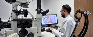
Have you ever wondered what exactly is doing the pathologist? They are very busy specialists who examine the tissue. But how does the whole process look like? Let's consider a simple example. We have a patient with some kind of small lump. We have no idea if it is a benign or malignant tumor. The only one that can solve this mystery is the pathologist. They make a decision based on the visual observation of the tissue from inside the lump under the microscope.
The tissue needed for evaluation can be taken either by biopsy or during the operation. Then, in the laboratory, it goes through a special process. It hardens and gets enveloped in a special substance, finally it becomes ready to be cut into thin layers that are then stored between two glass slides. Specimens prepared this way are ready for inspection with a microscope. Sometimes, the regular process is not enough. To enhance the probability of a good diagnosis additional slices are cut from the blocks and special staining (e.g. immunohistochemical) is used to augment visibility of extra features normally hidden in the tissue.
Many aspects of our modern life are digitalized, like online newspapers or mobile banking. This is also a case for healthcare and it is especially developing at a fast pace for pathology. Nowadays, it becomes digital pathology. Similarly to digital photography we can get a digital copy of a thin specimen, it has a fancy name of a virtual slide. It means not only that you can use computer instead of the microscope to observe the images, the possibility of processing the digital image of the tissue is now open. We can apply specially designed algorithms, filters and artificial intelligence for processing.
The typical evaluation done by pathologist directly via microscope is very time-consuming and tiresome. Moreover, the specialists have a lot of specimens to evaluate every day. It makes the human direct evaluation irreproducible in the exact same form. The evaluation process takes into account the features such as the architecture of the tissue sample, the intensity of staining as well as the amount of stained cell nuclei. Imagine counting hundreds of colorful objects every day in front of a microscope. This is the spot where the concept of this project takes initiative. The idea was to make the tissue evaluation easier by aiding the pathologist with task of counting the stained cell nuclei.
The software developed in this project deals with very big images, virtual slides, which required forming a strategy for dividing them into smaller parts and then accumulating the results. By introducing innovative algorithms for object detection and separation along with multiprocessing and application of artificial intelligence we managed to achieve satisfactory automation level of evaluation process. This gives a good perspective for evaluation of broad spectrum of stained tissue samples.
Utilization of our software can effectively increase the capability and efficiency of the diagnostic laboratory. One of the main advantages is the incredible speed of processing as well as reproducibility of the results. All this leads to make the process of diagnostics more efficient, reliable and consequently increase the number of cancer patients that recover (remission or survive).
Research project objectives/ Research hypothesis
The objective of this research is creating the supporting software to assist pathologist in evaluation of immunohistochemically stained tissue samples. Created technique will be tested on samples of breast cancer tissue stained with DAB&H (3,3' Diaminobenzidine&Haematoxylin) chosenfrom the tissue microarray. Developed method will be compared to the other known similar methods as well as to the manual evaluation of the pathologist.
Research project methodology
For the purpose of this research we intend to use the acknowledged methods of image processing and segmentation as well as our own innovative means. Apart from that, neural networks and techniques of parallel computing is planned to be used. To achieve segmentation of the immunopositive and immunonegative objects the variety of thresholding algorithms will be tested. We will focus on the locally adaptive thresholding methods where local threshold is calculated at every point of image while threshold value is based on the intensity of the pixel and its neighborhood. It seems to be the most appropriate approach in view of the fact that there are contrast fluctuations between objects of interest and background across image plane and from image to image. We already tested some methods and acquired good results. Although, the immunohistochemically stained tissue samples are acquired as 3-channel RGB images, we plan to separate specific dye from the image with the colour deconvolution algorithm. The conversion of images to the other colour model such as HSV or Lab will be considered as well.
Apart from the artificial images we will use experimental data collected from the tissue microarrays. To evaluate the whole sample size the method of selecting regions of interest will be developed next. We assume that the neural network will be able to make the decision upon chosen set of features such as texture, intensity variation, densitometry, etc, taken from the fragment of the image. The selected fragments are divided into parts which overlap, than parallel analysis is done, finally the algorithm eliminates unnecessary parts of results from the final objects maps and counting results.
In final stage the project the proposed method results are compared to the known methods results treated as ‘golden standard’. Because we try to achieve most accurate results that can be relied on, only the objects that our system is "sure" is automatically classified as positive or negative while other objects are presented to pathologist who by one click make decision. The system will be memorizing the manual grading so it could be upgraded manually or even improved interactively with every new case. To conclude this research the graphical user interface will be made so that nonprogrammer user can use this software.
Expected impact of the research project on the development of science, civilization and society
Immunohistochemically stained tissue samples are used by pathologists to establish the diagnosis, the prognosis and the treatment in various types of cancer. The evaluation process takes into account the amount of immunopositive objects and the architecture of the tissue sample. Such evaluation can be done by the experienced pathologist directly via microscope or from digital images of the samples. The human direct evaluation is irreproducible, time-consuming as well as intra- and inter-observer error prone. On the other hand automated methods, based on the digital image processing can improve the evaluation. The results are reproducible and they can become foundation of inter- and intra-laboratory unification in cut-offs and threshold levels. There are some more or less effective automatic and semi-automatic methods which extract immunopositive and immunonegative objects from images acquired from chosen by operator part of tissue sections but effective solutions developed for tissue microarrays are still not available.
The main problem is that there is no effective method that automatically selects regions of interest, which are areas where pathologist expects cancer cells without tissue not influenced by the disease and artifacts caused by sample preparation process. In this project the approach based on neural networks is proposed because of the power of this methodology. All this leads to make process of diagnostics more efficient and reliable and consequently more cancer patients overcome and recover (remission or survive).
Publications:
- Lukasz Roszkowiak, Anna Korzynska, Dorota Pijanowska; “Short survey: adaptive threshold methods used to segment immunonegative cells from simulated images of follicular lymphoma stained with 3,3'-Diaminobenzidine&Haematoxylin”, Proceedings of the 2015 Federated Conference on Computer Science and Information Systems (IEEE Annals of Computer Science and Information Systems), tom 5, str. 291-295 (10 pkt. MNSiW);
- Lukasz Roszkowiak, Anna Korzynska, Dorota Pijanowska; "Segmentacja immunonegatywnych jąder komórkowych w obrazach barwionych preparatów immunohistochemicznych raka sutka z użyciem DAB&H – analiza wstępna", Abstrakty KKBIB XIX, 2015, str. 189;
- Lukasz Roszkowiak, Anna Korzynska, Krzysztof Siemion, Dorota Pijanowska; „The Influence of Object Refining in Digital Pathology”; Image Processing and Communications Challenges 10. IP&C 2018. Advances in Intelligent Systems and Computing, vol 892. Springer, Cham;
- Lukasz Roszkowiak, Anna Korzynska, Carlos Lopez, Ramon Bosch, Marylene Lejeune, Jakub Zak, Krzysztof Siemion, Dorota Pijanowska; "Nuclei detection methods in DAB&H stained breast cancer biopsy images" (manuskrypt aktualnie w trakcie recenzji w The Journal of Histotechnology)
- Lukasz Roszkowiak, Anna Korzynska, Carlos Lopez, Ramon Bosch, Marylene Lejeune, Dorota Pijanowska; "New way to split the clusters based on distance transform in digital pathology" (manuskrypt aktualnie w trakcie recenzji w EURASIP Journal on Image and Video Processing – IF 1.534)
- "CHISEL: Framework for analysis of DAB&H stained breast cancer biopsy images", publikacja w trakcie przygotowania
Software:
Download from MathWorks FileExchange:
https://www.mathworks.com/matlabcentral/fileexchange/73365-chisel

